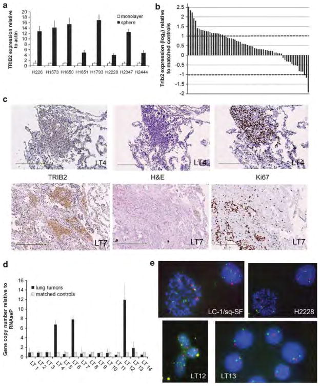
Figure 1.
TRIB2 is overexpressed and amplified in a subset of human lung tumors. (a) Expression of TRIB2 in eight NSCLC cell lines grown as monolayers (white bars) or under low-adherence conditions as spheres (black bars). Expression levels in spheres were normalized to actin and expressed relative to the values in monolayer cultures (P<0.01). (b) Microarray expression analysis of TRIB2 in 68 primary lung tumor samples. Data were normalized to the corresponding value from matched non-tumor samples and expressed as log2. Changes greater than two-fold were considered significant, indicated by the dashed lines. (c) Histology of human lung tumors stained for TRIB2 and Ki67 expression (in brown). Human lung tumor sample LT4 (upper panels) and LT7 (lower panels) stained with TRIB2, hematoxylin and eosin (H&E) and Ki67 to identify dividing cells. Scale bar is 250 μm. (d) Quantitative PCR of TRIB2 gene copy number based on a custom Taqman probe to the second intron. Results are normalized to RNAseP and expressed relative to matched non-tumor samples. Black bars represent gene copy numbers in tumor samples, gray bars represent the matched control tissues from each patient. (e) Fluorescence in situ hybridization analysis from lung cancer cell lines LC-1/sq-SF and H2228, and dissociated cells from human lung tumor samples LT12 and LT13. Probes for TRIB2 gene (green) and centromere of chromosome 2 (red) were used. Yellow spots represent non-specific background.

