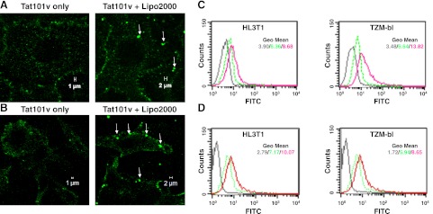
Figure 2.
Morphological characterization of fluorescent Tat proteins complexed to cationic liposomes. A) Binding of Tat101v without or with Lipo2000 (1:500) in HL3T1 cells after 20 min incubation. Lipo-Tat complexes are seen on the cell membrane (arrows). B) Binding of Tat101v without or with Lipo2000 (1:500) in TZM-bl cells after 20 min incubation. C) Membrane attachment of Tat-FITC complexed with Lipo2000 (1:200) or uncomplexed on HL3T1 and TZM-bl cells after 20 min incubation. D) Intracellular uptake of Tat-FITC with or without Lipo2000 (1:250) in HL3T1 and TZM-bl cells after incubation for 2 h and trysinization for 15 min. Colors in graphs of flow cytometry: dark gray, untreated cells; green, Tat-FITC only; red, Tat-FITC complexed with Lipo2000. View in images: ×630.

