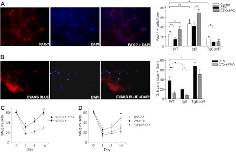
Figure 2.
EPO enhanced recovery in CTX-induced muscle injury. A) Three groups of mice each (4 wk in age; sex matched; n=6) from TgEpoR, tg6, and WT littermate mice were treated with 100 μl PBS (control; open bars), PBS containing CTX (10 μM; solid bars) and PBS containing CTX + EPO (3000 U/kg; shaded bars) by injection into the gastrocnemius muscle. Muscles were harvested 3 d after injection, and paraffin sections were stained for Pax-7 (red) and DAPI (blue). Representative sections are shown (left panels), and Pax7+ cells were quantified (right panel). B) Mice were treated as described in A, and at 24 h prior to tissue collection, Evans blue dye solution (1%) was injected intraperitoneally. Muscles were harvested and stained for Evans blue dye (red) and DAPI (blue). Representative sections are shown (left panels), and percentages of Evans blue-positive fibers were quantified and compared for CTX treatment without (solid bars) and with EPO treatment (shaded bars). C) Muscle tension to rupture determined for dissected gastrocnemius muscle was used to assess muscle regeneration before (d 0) or after CTX treatment (d 1, 3, and 14) in WT mice without (solid line) and with EPO treatment (3000 U/kg; dashed line). Mice were 4 wk of age (female; n=6). D) Muscle tension to rupture of the isolated gastrocnemius muscle was determined for TgEpoR (open triangles; dashed-dotted line), tg6 (open circles; solid line) and WT littermate mice (solid circles; dashed line) 4 wk in age (sex matched, n=6) before (d 0) or after CDX treatment (d 1, 3, and 14). *P < 0.05; **P < 0.01.

