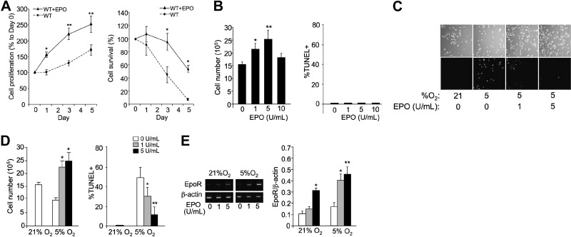
Figure 4.
EPO protects myoblasts from 5% O2-induced apoptosis. A) Cell proliferation was determined for primary myoblasts from gastrocnemius muscle isolated from 4-wk-old WT mice that were cultured without (circles; dashed line) and with EPO (5 U/ml; triangles; solid line) for 5 d (left panel). Primary myoblast cultures were also exposed to 5% O2 for 5 d; percentage of surviving cells compared to d 0 is shown (right panel). B) Proliferation (left panel) and TUNEL assay (right panel) of C2C12 cells seeded at 1 × 10⁶ cells/well in 6-well pales and treated with EPO (0, 1, 5, and 10 U/ml) for 24 h were determined. C) C2C12 cells seeded at 1 × 10⁶ cells/well in 6-well plates, treated with EPO (0, 1, and 5 U/ml), and cultured at 5% O2 for 24 h were compared with cells cultured at 21% O2. D) Quantification of cell numbers (left panel) and percentage of TUNEL+ cells (right panel) from C. E) EpoR and β-actin mRNA expression in C2C12 cells cultured at 21 and 5% O2 and without and with EPO treatment (5 U/ml) was determined by quantitative real-time RT-PCR. EpoR expression is normalized to β-actin as the internal control. **P < 0.01.

