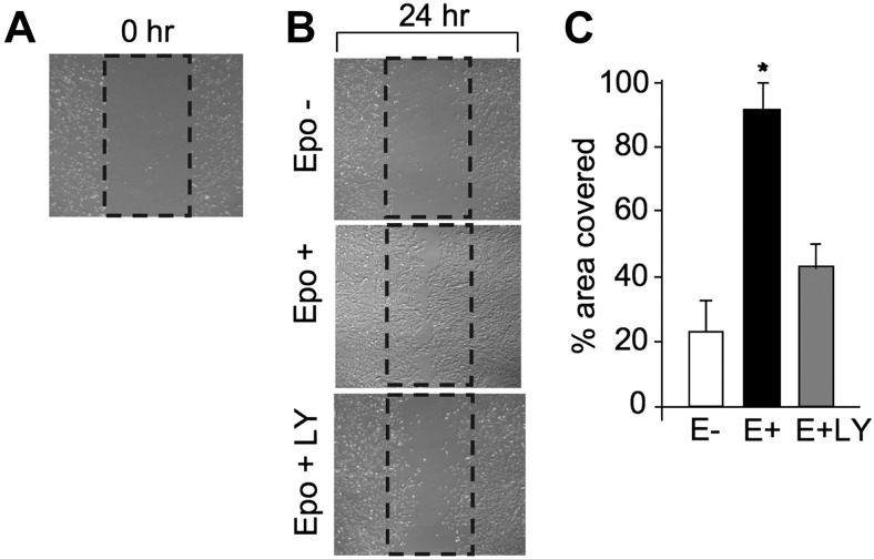
Figure 5.
Cell scrape-wound assay. A) C2C12 cells were cultured in growth medium to ∼80% confluence for the in vitro scratch assay. Percentage of area covered by myoblasts growing back into the scraped area after 24 h was monitored. B, C) Cells were treated without (open bar) and with EPO (5 U/ml; solid bar) and EPO plus LY-294002 (LY; 50 μM; shaded bar) after the cells were scraped. After 24 h, images of the scraped area were captured by Nikon phase-contrast microscope (B) and the percentage of the scraped area covered by myoblasts was determined by the Image Pro program (C). *P < 0.05.

