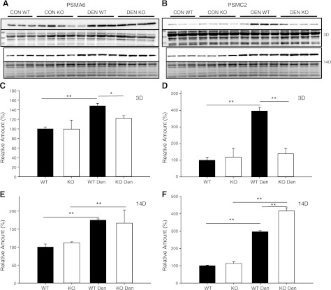
Figure 4.
Protein expression of 19S and 20S proteasome subunits in control (con) and denervated (den) muscle. A, B) Western blots of lysates from the TS of WT (n=3–4) and MuRF1-KO (n=4) mice following 3 and 14 d of denervation. Immunoblots for PSMA6 (α1, 20S subunit; A, top panel) and Ponceau staining (A, bottom panel) and PSMC2 (Rpt1, 19S subunit; B, top panel) and Ponceau staining (B, bottom panel). C–F) Quantification of immunoblots from WT (solid bars) and MuRF1-KO (open bars) mice; data are expressed as means ± sd. Relative amounts (expressed as a percentage of WT con) of PSMA6 protein (C, E) or PSMC2 protein (D, F) in TS after 3 d (C, D) or 14 d (E, F) of denervation. *P < 0.05, **P < 0.001.

