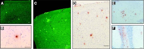
Figure 3.
Histochemical and immunocytochemical detection amyloid deposits. A, B) Only few amyloid deposits were detected in the neocortex of young mice by Th-S (A) or immunohistochemistry (B). C–E) As animals aged, abundant parenchymal Aβ deposition was observed in the cerebral cortex (C, D) and hippocampus (E). In addition to amyloid plaques, leptomeninges also showed the presence of fibrillar Aβ (C, D). F) In the cerebellum, amyloid deposition was observed in leptomeningeal vessels and in the parenchyma. Brain sections from 5 (A), 6 (B), 15 (C, D), and 20-mo-old (E, F) APP YAC × Psen1-L166P (+/+) mice. Th-S (A, C). Immunohistochemistry using Ab 4G8 (B, D, F) and 21F12 (E). Scale bars = 100 μm (A, C, D); 50 μm (B, E, F).

