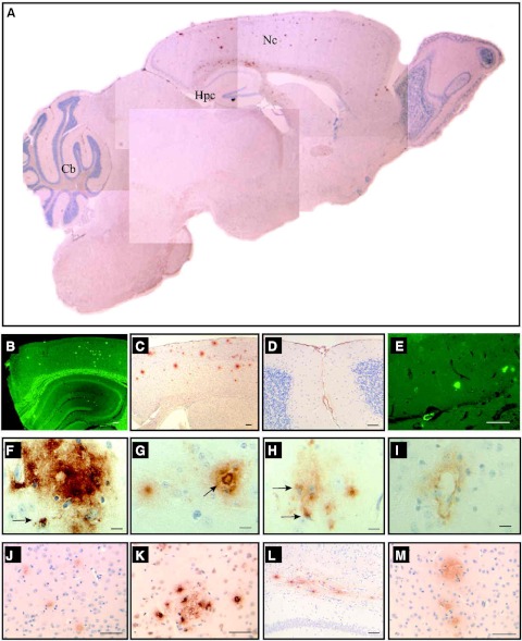
Figure 4.
Distribution of amyloid deposits in APP YAC × Psen1-L166P mice. Sagittal section of an APP YAC × Psen1-L166P (+/+) mouse (A) shows the regional distribution of amyloid deposits in the brain. Numerous amyloid deposits were detected in the neocortex (Nc) by immunohistochemistry using antibodies against different epitopes of the Aβ peptide (A, C, F–H, J, K, M). In the cerebellum (Cb), amyloid deposition was observed in pial (leptomeningeal) vessels (A, D). In the hippocampus (Hpc), Th-S fluorescent plaques were observed in addition to amyloid deposits in the walls of vessels of the hippocampal fissure (A, E). Severe amyloid deposition is observed in the corpus callosum (L). In the neocortex, different varieties of amyloid deposits were observed, including diffuse (F, G), dense-cored (arrow, G), and vascular (I). Some deposits were clearly intracellular (arrows, F, H). Brain sections from an APP YAC × Psen1-L166P (+/+) mouse (15-mo-old). Immunohistochemistry using Abs 4G8 (A, D, F–I), 6E10 (C, L), 10D5 (J), 21F12 (K), and 3D6 (M). Th-S (B, E). Scale bars = 100 μm (B, C); 50 μm (D, E, J–M); 10 μm (F–I).

