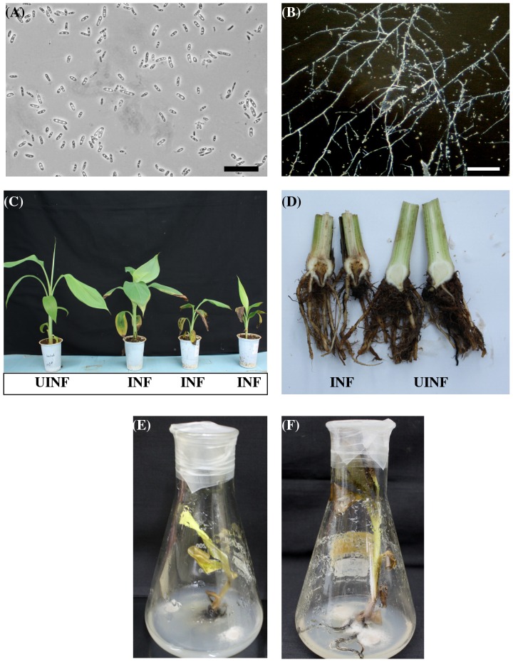
Figure 3. Identification and pathogenicity testing of the Foc isolate.
Foc oval shaped spores (A) and mycelium (B) as observed under microscope. Bars correspond to 25 µm (C) Hardened plants of banana cv. Rasthali showing initial symptoms of Foc race 1 infection 3 weeks post inoculation (INF). An uninfected control banana plant is shown on the extreme left (UINF). (D) Necrotic lesions developed inside the corm subsequent to Foc race 1 inoculation on banana cv. Rasthali (left). An uninfected control banana plant is shown on the right. Foc infected untransformed control plants showing disease symptoms after 2 weeks (E) and 4 weeks (F). Mycelium is seen covering the roots of the infected plant.

