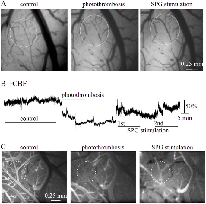
Figure 3. SPG stimulation in the RB-treated cortex.
A, Brain surface images in a typical experiment before (left), and after (middle) photothrombosis and during SPG stimulation (2 mA, 500 µs, right). Note vasodilation and reperfusion of the thrombosed vessels (circled). B, Laser-Doppler recording in the same experiment as in (A), showing reduced rCBF during photothrombosis, which was partially reversible during SPG stimulation. C, Fluorescent angiography from a different rat before (left), after photothrombosis (middle) and during SPG stimulation (2 mA, 500 µs, right).

