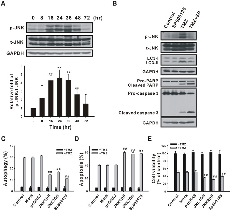
Figure 9. Participation of JNK in TMZ-induced autophagy.
(A) U87 MG cells were treated with 400 µM TMZ for the indicated time periods and then analyzed using immunoblotting with anti-p-JNK and anti-t-JNK antibodies. The amount of protein applied in each lane was normalized by t-JNK internal control. (B) U87 MG cells were pretreated with 10 µM SP600125 for 1 h, followed by treatment with TMZ for 36 h (for analysis of JNK and LC3) or 72 h (for analysis of PARP and caspase 3). Cell lysates were analyzed by immunoblotting with anti-p-JNK, anti-t-JNK, anti-LC3, anti-PARP, anti-caspase 3, and anti-GAPDH antibodies. GAPDH and t-JNK were used as internal controls to normalize the amount of protein applied in each lane. U87 MG cells were transfected with 1 µg of the pcDNA3, JNK1DN, or JNK2DN plasmid for 48 h, or pretreated with 10 µM SP600125 for 1 h, followed by treatment with 400 µM TMZ for another 72 h to determine percentages of cells undergoing autophagy (C) and apoptosis (D) as well as cell viability (E). Results are presented as the mean ± SD. t-JNK, total JNK; p-JNK, phosphorylated JNK. **p<0.01 vs. each respective control. *p<0.05, **p<0.01 vs. each respective TMZ group.

