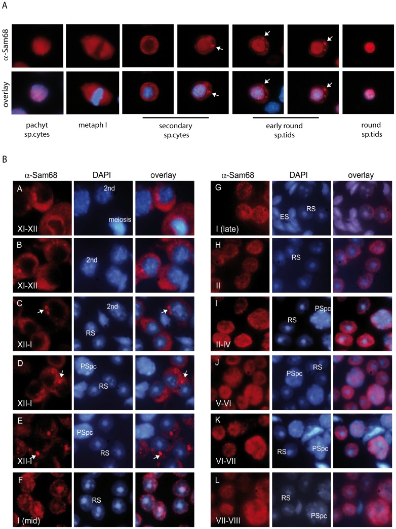
Figure 1. SAM68 accumulates in a perinuclear organelle in secondary spermatocytes and early round spermatids.
(A) Purified male germ cells were stained with an anti-SAM68 antibody (red) and co-stained with Hoechst (blue) to detect nuclei and to identify cell stages by nuclear morphology. In secondary spermatocytes and early round spermatids SAM68 accumulates into a granule (white arrows) resembling the chromatoid body. (B) Stage specific localization of SAM68 during spermatogenesis. Squashes of male germ cells from seminiferous tubules at different stages of spermatogenesis show that SAM68 (red) localizes in the cytoplasm and was enriched in perinuclear granules (arrows) in meiotic spermatocytes from stage XII tubules and in early round spermatids from stages XII and I (A–E). In late stage I spermatids and from stage II through VIII, SAM68 was predominantly nuclear (F–L). Cells were co-stained with DAPI to detect nuclei. 2nd = secondary spermatocytes; RS = round spermatid; PSpc = pachytene spermatocyte; ES = elongated spermatid.

