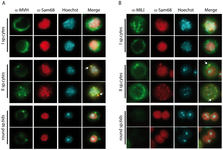
Figure 2. Co-localization of SAM68 with MVH and MILI in male germ cells.
(A) Isolated male germ cells were co-stained with an anti-SAM68 antibody (red), an anti-MVH antibody (green) and with Hoechst (blue) to detect nuclei. SAM68 and MVH partially co-localize in the CB of secondary spermatocytes (arrows), while in primary spermatocytes SAM68 is nuclear and MVH is cytoplasmic, and in round spermatids SAM68 is nuclear and MVH is predominantly localized in the CB. (B) Isolated germ cells were analysed by immunofluorescence using the anti-SAM68 antibody (red) and the anti-MILI antibody (green). Nuclei were stained with Hoechst (blue) to identify cell stages by nuclear morphology. In primary spermatocytes SAM68 localizes in the nucleus, while MILI is cytoplasmic; in round spermatids SAM68 is nuclear and MILI is absent. The localization of the two proteins partially overlaps only in the CB of secondary spermatocytes.

