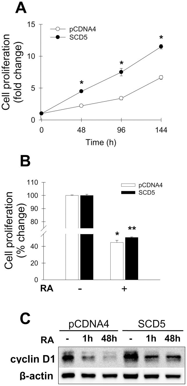
Figure 5. Expression of human SCD5 in Neuro2a cells increases cell proliferation. Effect of retinoic acid.

Neuro2a cells stably transfected with human SCD5 cDNA (SCD5 cells) or with empty plasmid (pCDNA4 cells) were seeded in 12-well plates and grown for different time points up to 120 h (A). At each time point, cell proliferation was determined by Crystal violet assay as described in Materials and Methods. B, SCD5-expressing Neuro2a cells and their controls were incubated with 10 µM retinoic acid or DMSO vehicle for 48 h. Cell growth was estimated by Crystal violet staining method. Values represent the mean ± S.D. of triplicate determinations. *, p<0.05 or less vs control, by Student's t test. C, Western blot determination of cyclin D1 and β-actin levels in control (pCDNA4) and SCD5-expressing Neuro-2a cells incubated in serum-free DMEM with 0.1% BSA, in presence or absence of 10µM retinoic acid (RA), for 1 h or 48 h.
