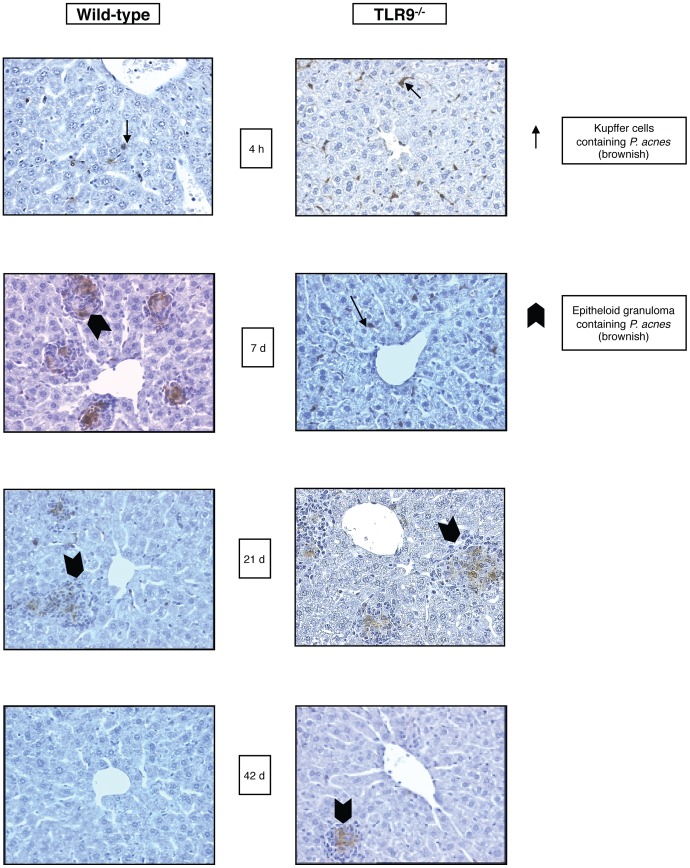
Figure 1. Persistence of P. acnes in livers of WT and TLR9−/− mice.
Groups of five WT and TLR9−/− mice were treated with heat-killed P. acnes (100 µg/g b.w.) i.v. and livers were removed at different time points after treatment. Immunohisto-chemical staining of liver sections using P. acnes specific antiserum shows brown P. acnes-specific staining within granulocytes, sinusoidal lining cells and/or granulomas. Left panel: liver of WT mice; right panel: liver of TLR9−/− mice. One representative slide per group of five animals is shown.

