Figure 1.
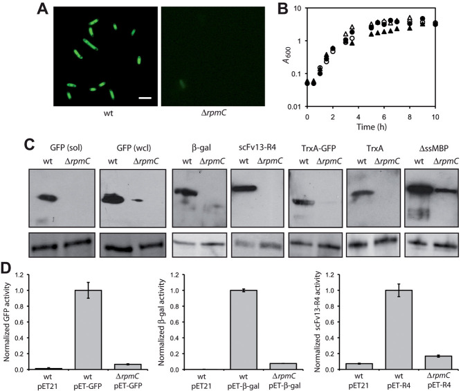
Expression of recombinant proteins in L29-deficient cells. (A) Fluorescence microscopy and (B) cell growth of wt and ΔrpmC::kan cells expressing GFP. Scale bar, 1 μM. Growth curves show induced (closed) and uninduced (open) cultures of wt (triangle) or ΔrpmC::kan (circle) cells. Data show the average of three independent experimental repeats and the SEM for these data was less than 5%. (C) Western blot analysis of whole cell lysates (wcl) isolated from wt and ΔrpmC::kan cells expressing different target proteins as indicated. For GFP, the soluble (sol) fraction is also shown. An equivalent number of cells was loaded in each lane. GroEL served as a loading control (lower panels). (D) Activity of GFP, β-gal and scFv13-R4 measured in whole cell lysates prepared from wt and ΔrpmC::kan cells carrying an empty vector control (pET) or a pET vector with the indicated protein. GFP activity was measured by FACS and cells were ungated, β-gal activity was measured using the Miller assay, and scFv13-R4 activity was determined by ELISA. All values were normalized to the activity measured in wt cells. An equivalent number of cells was assayed in each case. Data show the average of at least three independent experimental repeats and the error bars represent the SEM.

