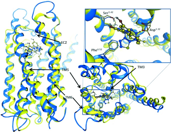
Figure 7.

Two orthogonal views of the dopamine D1 (blue) and D2 (yellow) receptor models together with the corresponding full agonists (R)-2-OH-NPA (blue) and SKF89626 (yellow) present in their binding sites. The typical monoaminergic key interacting amino acid residues are shown explicitly. The structures differ particularly in the second and the third extracellular loops (EC2 and EC3), but also in the transmembrane (TM) region, where important interacting amino acids are positioned.
