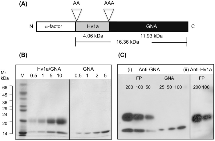
Figure 1. Protein production and purification.
(A) Schematic of construct encoding Hv1a/GNA showing predicted molecular masses of Hv1a and GNA as well as the total mass of the Hv1a/GNA fusion protein including the tri-alanine linker region and the additional two alanine residues at the N-terminus. (B) Coomassie blue stained SDS-PAGE gel (17.5% acrylamide) of recombinant Hv1a/GNA and GNA following purification by hydrophobic interaction and gel filtration chromatography. The approximate loading of protein (µg) is indicated above each lane, while the lane marked “M” contains molecular weight standards (Sigma SDS-7). (C) Composite of Western blots of recombinant proteins using (i) anti-GNA and (ii) anti-Hv1a antibodies. The approximate protein loading (ng) is denoted above each lane. FP denotes Hv1a/GNA fusion protein.

