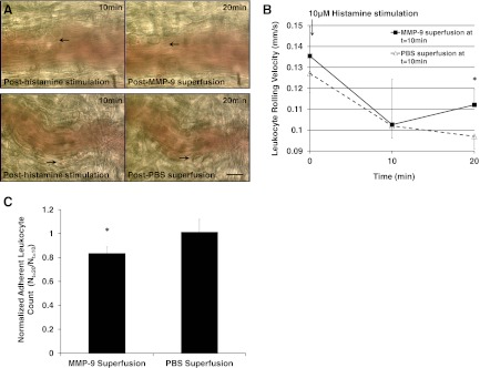
Figure 5. Positive control experiment: 10 min superfusion of MMP-9 (1.63 nM) or 1× PBS (as negative control) to WKY postcapillary mesenteric venules (n=3 rats) post-10 min histamine (10 μM) stimulation.
(A) Representative images of postcapillary venules subject to MMP-9 or PBS superfusion with adherent leukocytes indicated by black arrows at t = 10 min and 20 min. (B) Average leukocyte rolling velocity at t = 0, 10, and 20 min. We analyzed n = 20 leukocytes and averaged their rolling velocities along an 80 μm venular segment/rat. One-tailed Student's t test: *P < 0.05 MMP-9 versus PBS superfusion at t = 20 min. (C) Average adherent leukocyte count (Nt=20 min) after MMP-9 or PBS superfusion normalized to leukocyte count (Nt=10 min) before MMP-9 or PBS superfusion. *P < 0.05 MMP-9 versus PBS superfusion.

