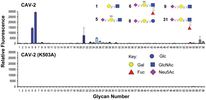
Fig. 2.
Glycan array analysis of the CAV-2 fiber knob protein binding to a printed sialoside library. (Top) The adenoviral capsid protein (50 µg/mL) strongly recognizes the two 6′-sulfated α2,3-linked sialosides 5 and 6. (Bottom) Site-directed mutagenesis of lysine 503 to an alanine abrogated binding to the printed glycans (see Supplementary data for the complete list of glycans, Supplementary data, Table S2 and Figure S1 for glycan array analysis at higher protein concentrations).

