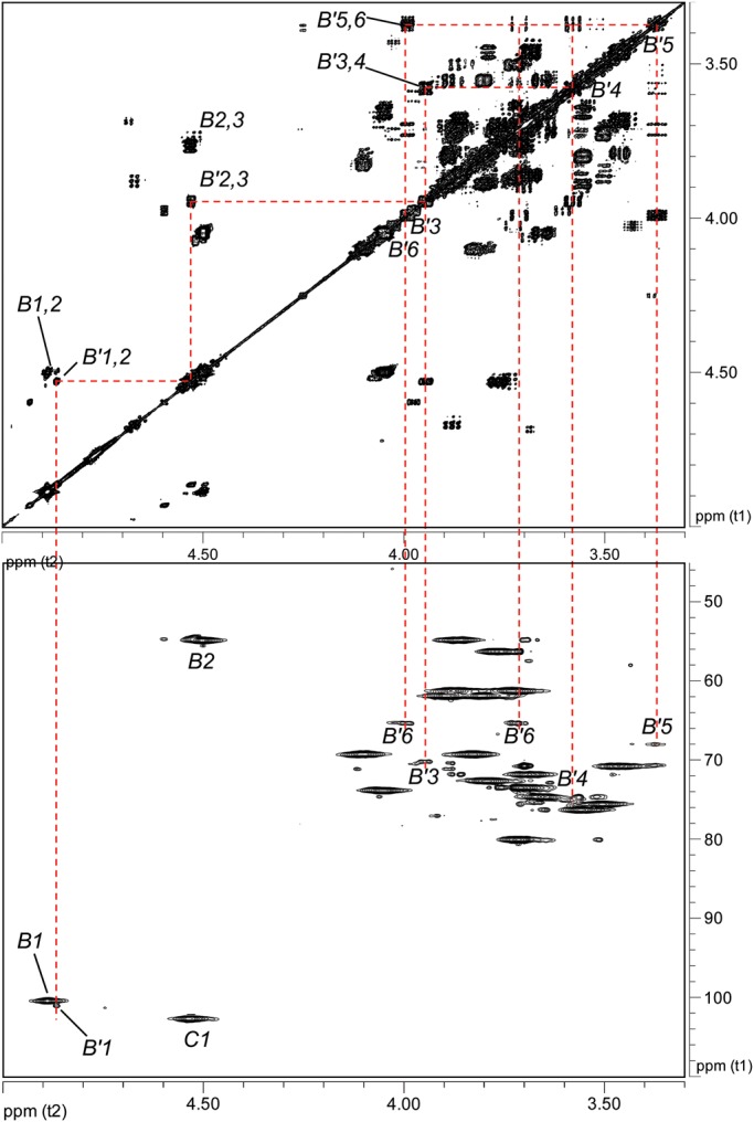
Fig. 6.

Partial 1H–1H COSY and 1H–13C HSQC spectra of the Ba684 SCWP defining the terminal pyruvylated-ManNAc (residue B′). Top panel, COSY; bottom panel, HSQC. The spectra, displayed at high magnification, allow the visualization of the weak signals arising from the B′ spin system. This residue occurs at only one location, the terminal non-reducing end of the SCWP.
