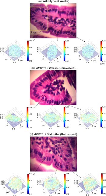
Fig. 2.
Representative regular bright-field images of H&E stained epithelial cell nuclei and corresponding structure-derived nuclear optical path length (OPL) maps for three selected cells derived from (a) wild-type mouse (six weeks), (b) histologically normal appearing cells (uninvolved cells) from six weeks mouse, and (c) histologically normal appearing cells (uninvolved cells) from 4.5 months mouse. The marked area in part (a) shows the representative columnar shaped epithelial cells having similar morphological features.

