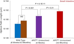
Fig. 3.
Statistical analysis of the nuclear nano-morphology marker characterized by structure-derived optical path length (OPL) from the cell nuclei of wild-type mice and histologically normal (i.e., uninvolved) appearing intestinal epithelial cell nuclei from gender and age-matched mice at six weeks and 4.5 months. The nuclear OPL exhibits a progressive increase with the development of carcinogenesis. Approximately 50 to 60 cell nuclei were analyzed from each mouse.

