
Fig. 2.
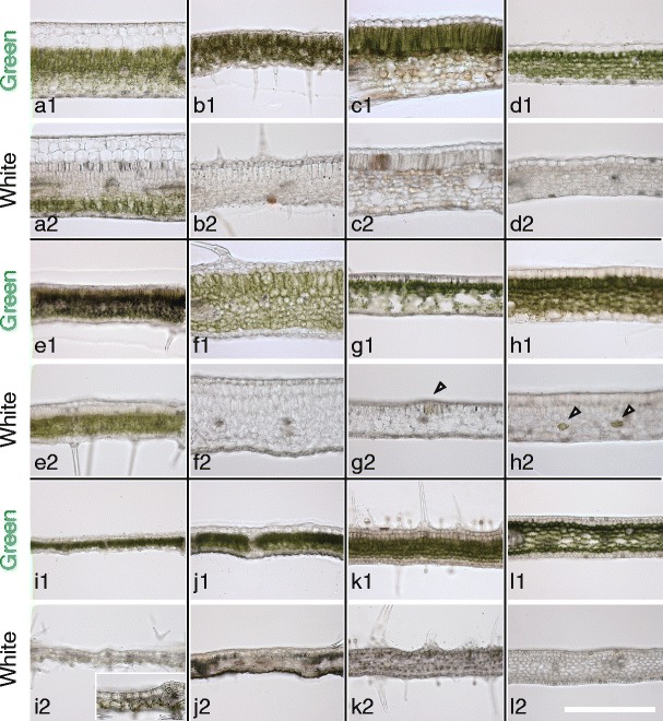
Cross sections of variegated leaves used in this study. a1/a2 Schefflera arboricola b1/b2 Solenostemon scutellarioides; c1/c2 Impatiens hybrids; d1/d2 Hosta sp.; e1/e2 Ipomea nil; f1/f2 Felicia amelloides; g1/g2 Hedera canariensis; h1/h2 Epipremnum aureum; i1/i2 Abutilon variegatum; j1/j2 Fragaria × ananassa; k1/k2 Pelargonium zonale; l1/l2 Dracaena sanderiana. Cross sections of green (a1, b1, c1, d1, e1, f1, g1, h1, i1, j1, k1, l1) and white (a2, b2, c2, d2, e2, f2, g2, h2, i2, j2, k2, l2) sectors are shown. Arrowheads in g2 and h2 show green cells in white sectors. Inset in i2 is a higher-magnification image showing the green color of abaxial epidermal cells. Scale bar in l2 represents 500 μm