
Fig. 4.
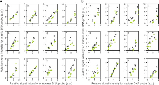
Comparison of organelle DNA levels in green and white sectors. A Comparison of plastid DNA level. B Comparison of mitochondrial DNA level. Total DNAs were extracted from green and white sectors of the same leaves and dot-blotted to nylon membrane as dilution series (75, 150, and 300 ng/dot or 50, 100, and 200 ng/dot). Three sheets of membrane with identical contents were prepared, each of which was hybridized with a single probe that was specific to a plastid, a mitochondrial, or a nuclear DNA sequence. For each plant species, hybridization experiments were repeated three times using three different sets of DNA samples. Intensities of hybridization signals were measured and transformed to relative values, where, for each plant species, each probe and each white/green pair, the average of the signal intensities from green and white sector samples at middle dose (100 or 150 ng/dot) was arbitrarily set at 1. Finally, the relative signal intensities for plastid DNA probe (A) and mitochondrial DNA probe (B) were plotted against the relative signal intensities for nuclear DNA probe. Green and white symbols indicate data from green and white sectors, respectively. Different figures of symbols (circles, triangles, and squares) indicate results from different set of leaves. Data with extremely low signals (i.e., those without linear dose–signal relationships) were excluded from the analyses. a Schefflera arboricola; b Solenostemon scutellarioides; c Impatiens hybrids; d Hosta sp.; e Ipomea nil; f Felicia amelloides; g Hedera canariensis; h Epipremnum aureum; i Abutilon variegatum; j Fragaria × ananassa; k Pelargonium zonale; l Dracaena sanderiana. For results of statistical analyses, see Table 3