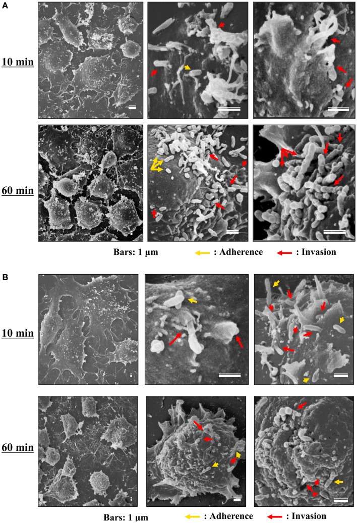
Figure 3.
Scanning electron microscopy of cells infected with TGR and SFGR. (A) AeAl2 cells infected with R. typhi at 10 and 60 min after infection. (B) AeAl2 cells infected with R. japonica at 10 and 60 min after infection. Successful adherence to and invasion of AeAl2 insect cells was achieved by both TGR and SFGR soon after their inoculation. The yellow and red arrows indicate adherent and invading rickettsiae, respectively.

