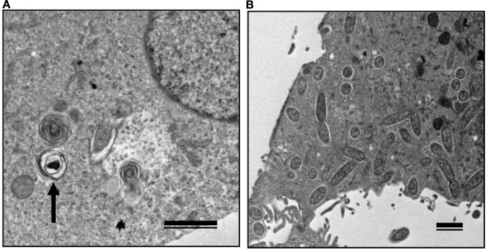
Figure 6.
Transmission electron microscopy of Vero cells infected with non-pathogenic and pathogenic SFGR. (A) Vero cells infected with R. montanensis alone was observed at 7 days after infection. An arrow marks a degenerating rickettsia in an autophagosome-like vacuole. (B) R. montanensis-infected cells were superinfected with R. japonica on day three of infection and observed at 7 days after the first infection. Many free rickettsiae around 1 μm in length surrounded by halos and those in the course of binary fission were seen in the cytoplasm. Bars, 1 μm.

