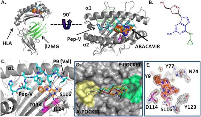
Fig. 3.
Crystal structure of the abacavir–peptide–MHC complex solved to a resolution limit of 2.0 Å reveals intermolecular contacts within the antigen-binding cleft. (A) Cartoon diagram of HLA-B*57:01 in gray. The peptide HSITYLLPV is shown in cyan carbons. Abacavir is shown as spheres, orange for carbon, blue for nitrogen and red for oxygen. (B) Chemical structure of abacavir, with the cyclopropyl moiety shown in green, the purine core in blue, and the hydroxymethyl cyclopentene moiety in red. (C) Abacavir forms H bond interactions (black dashes) with both the peptide and HLA-B*57:01. The residues that distinguish the abacavir-sensitive allele HLA-B*57:01 from abacavir-insensitive HLA-B*57:03 are shown in magenta for carbon, blue for nitrogen, and red for oxygen. (D) Abacavir binding in the F pocket does not alter the peptide conformation compared with other peptide/HLA-B complexes. A cartoon representation of peptide in the crystal structure complexed to abacavir and HLA-B*57:01 is shown in cyan (HSITYLLPV; PDB ID: 3UPR). A 9-mer self peptide (LSSPVTKSF) complexed to HLA-B*57:01 (PDB ID: 2RFX (2) is shown in red, the 8-mer peptide epitope HIV1 Nef 75–82 (VPLRPMTY) bound to HLA-B*35:01 (PDB ID: 1A1N) (40) is shown in pink, a 9-mer EBV peptide (FLRGRAYGL) complexed to HLA-B8 (PDB ID: 1MI5) (41) is shown in green, and the 11-mer EBV peptide HPVGEADYFEY complexed to HLA-B*35:01 (PDB ID: 3MV9) (42) is shown in yellow. The molecular surface of HLA-B*57:01 from 3UPR is shown in gray. The F pocket residues (9) are colored green, and the A pocket is yellow. (E) Experimental electron density corresponding to abacavir in an Fo-Fc difference map contoured at 3.5σ (red mesh) after molecular replacement. Gray mesh depicts the final 2Fo-Fc electron density map of abacavir in the antigen-binding cleft of HLA-B*57:01 (contour level, 1.5σ). H bond interactions between abacavir and HLA-B*57:01 are shown as yellow dashed lines. The residues that distinguish the abacavir-sensitive allele HLA-B*57:01 from abacavir-insensitive HLA-B*57:03 are shown in magenta for carbon, blue for nitrogen, and red for oxygen.

