Fig. 3.
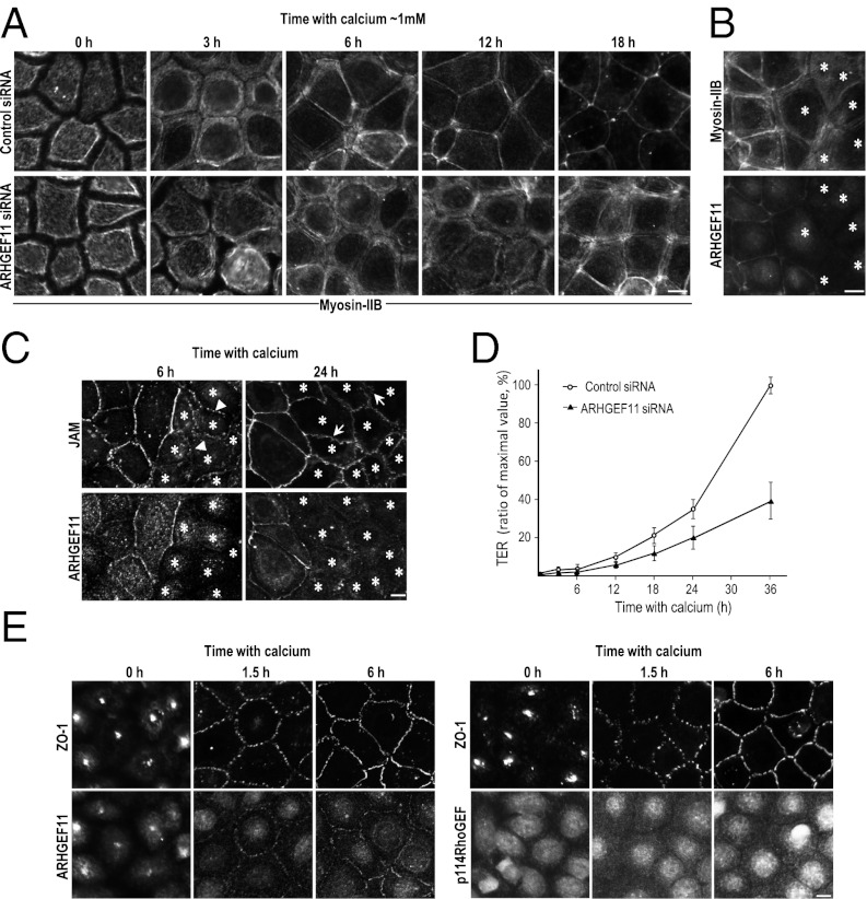
ARHGEF11 is implicated in junction assembly and epithelial barrier formation. (A) EpH4 cells transfected with control or ARHGEF11-specific siRNA were cultured in low-calcium medium overnight. Junction assembly was initiated by reintroducing normal calcium medium. Cells were fixed at the indicated times and immunostained for myosin-IIB. (B and C) To further verify the effect of ARHGEF11 depletion on PJARs or TJs, a calcium switch assay was conducted using coculture of control and ARHGEF11-depleted cells. Cells were stained for myosin-IIB ∼9 h after the calcium switch (B), and stained for JAM-A ∼6 h or ∼24 h after the calcium switch (C). Arrowheads and arrows indicate spot-like junctions and undulating appearance, respectively. Asterisks indicate ARHGEF11-depleted cells. (D) Establishment of the epithelial barrier was monitored by measuring the TER at the indicated times (data presented as means ± SD; n = 3). The y axis is the percentage of maximal TER measured in this experiment. (E) EpH4 cells were fixed at the indicated times after being returned to normal calcium, and the localization of ARHGEF11 or p114RhoGEF was compared with that of ZO-1. (Scale bars: 10 μm.)

