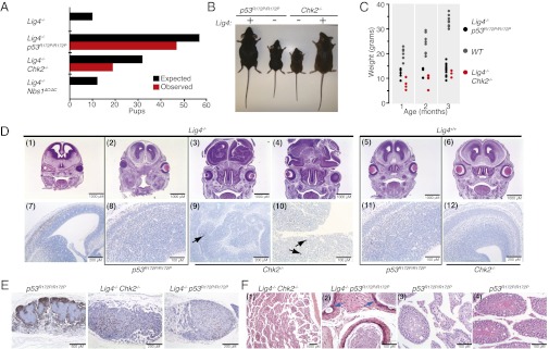
Fig. 2.
Apoptotic defects are sufficient to rescue the viability of Lig4-deficient animals. (A) Graphical representation of expected vs. observed number of pups based on Mendelian inheritance of alleles. Details of breeding schemes and numbers are provided in Table S8. (B) Representative images of 2-mo-old mice of the indicated genotypes. (C) Analysis of changes in pup weight of the indicated genotypes over 3 mo. (D) Immunohistochemical analysis of brain morphology in E13.5 embryos of the indicated genotypes. (Upper) H&E staining. (Lower) Cleaved caspase-3 staining. Arrows in panels 9 and 10 indicate rare clusters of caspase 3 positive cells. (E) B220 staining of mesenteric lymph nodes. (Left) Normal mesenteric lymph node of a p53R172P/R172P mouse. (Center and Right) Marked lymphoid atrophy/hypoplasia observed in all Lig4-null mice; genotypes are indicated. (F) Testicular pathology observed in 4/4 Lig4−/− Chk2−/− and 9/15 Lig4−/− p53R172P/R172P mice. (1) Degeneration of seminiferous tubules observed in Lig4−/− Chk2−/− mice between 3.2 and 7.1 (shown) mo. (2) Cross-section of 2.2-mo-old Lig4−/− p53R172P/R172P epididymis showing pleomorphic epithelial cells with karyomegaly (blue arrows) and vacuoles. (3) Variable loss of spermatogonia and primary and secondary spermatocytes observed in seminiferous tubules of the testes in a p53R172P/R172P mouse (age 14.8 mo). (4) Normal testes pathology in a p53R172P/R172P mouse (age 11.3 mo).

