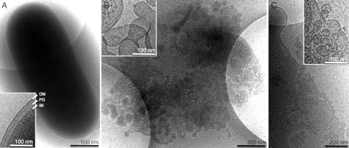
Fig. 3.
Cryo-EM of fyuA+ E. coli cells treated with the hybrid lysin. Electron micrographs of fyuA+ E. coli cells plunge-frozen on holey-carbon mesh coated sample grids. Cells were incubated with (A) no treatment (control) or (B and C) 100 μg/mL hybrid lysin for 10 min before being plunge-frozen. Extensive membrane breakdown of the treated cell in (B) is apparent. As a result, the cell is weakened and, consequently, spreads and flattens in the thin film of buffer while largely preserving its 3D surface area. As a result, it projects a larger 2D area in the micrograph. Also, as it is so much thinner, the membrane vesiculation is evident all over the cell, not just around the edges. The insets show typical regions of the corresponding cell envelopes at higher magnification. The intact inner membrane (IM), peptidoglycan layer (PG), and outer membrane (OM) are indicated and labeled in (A). Characteristic vesiculation of inner and outer cell membranes is shown in (B) and (C).

