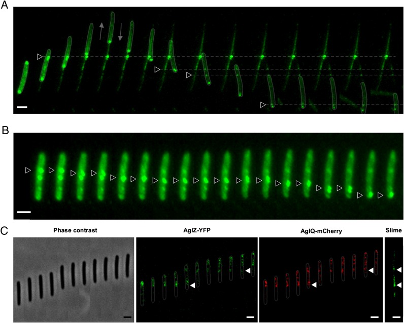
Fig. 4.
Slime is deposited by the Agl/Glt motility complexes. (A) ConA staining of a moving cell and its resulting slime trail at different times. Note that the cell changes direction after t5 (2 min). Triangular arrows point to conspicuous bright dots that appear at the leading cell pole and remained fixed relative to the substratum before they eventually became deposited on the substratum (for animation, see Movie S4). Fluorescent micrographs were taken every 30 s. (Scale bar, 1 μm.) (B) ConA clusters are transported down the cell body in immobile cells. Two ConA bright clusters (black triangles) are shown to move down the cell axis. Pictures were taken every 15 s. (Scale bar, 1 μm.) (C) Slime patches are deposited where the Agl/Glt machinery assembles. Time lapse of a cell expressing both AglZ-YFP and AglQ-mCherry is shown. Phase contrast and corresponding YFP and mCherry micrographs are shown. Slime was stained with ConA after the cell left the positions shown on the Left. Triangular arrows point to fixed AglZ- and AglQ-bright motility complexes at positions where conspicuous slime patches were deposited. Fluorescent micrographs were taken every 15 s. (Scale bar, 1 μm.)

