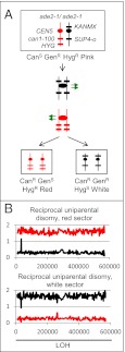
Fig. 3.
Diagnosis of RUD. (A) Pattern of marker segregation resulting from a RUD event. The chromosomes are depicted in the same way as in Fig. 2A. In the RUD event, unlike the reciprocal crossover shown in Fig. 2A, the cells in the red sector are geneticin-sensitive. (B) Analysis of genomic DNA isolated from red and white sectors from a RUD sectored colony [SLA11.7(43)]. Genomic DNA was purified from each sector of a canavanine-resistant red/white-sectored colony and examined as described for Fig. 2B. The red sector exhibits the hybridization patterns consistent with UPD for the W303a-derived chromosome, and the white sector has the patterns expected for UPD for the YJM789-derived chromosome.

