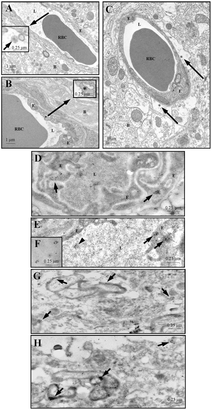
Figure 2. Panels A and B: HIV particles (arrows) in the lumen (panel A) and in an endothelial cell (panel B) of blood vessels in mouse cerebral cortex.
The boxed regions show higher magnification images. Panel C: HIV (arrows) have traversed the endothelium of a blood vessel in mouse brain cerebral cortex and gained access the perivascular space. Panels D–F: Colloidal gold labeled HIV in the lumen (arrowhead in panel E, and panel F) and in endothelial cells (arrows in panels D and E) of mouse cerebral cortex capillaries. Panels G and H: HIV associated with myelin sheaths (arrows) of nerve axons in mouse cerebral cortex. Thin sections were incubated with HIV-1SF2 gp120 antiserum diluted 1:100 and followed, after washing, by incubation in protein A-10 nm gold diluted 1∶100. B – brain parenchyma, E – endothelium, L – capillary lumen, RBC – red blood cell.

