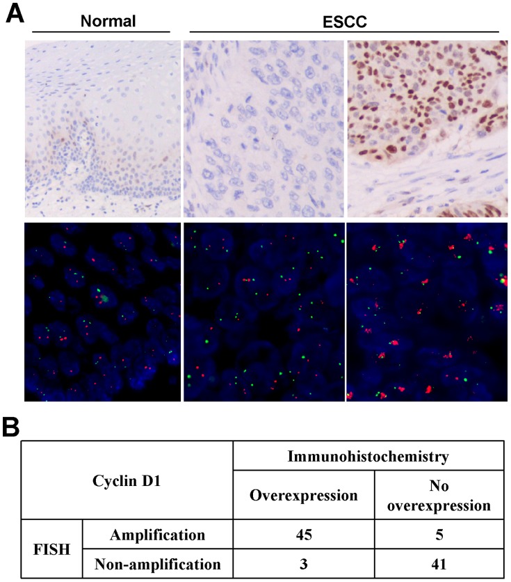
Figure 5. Representative immunohistochemical staining and FISH analysis.
. A, Top panels, immunohistochemical staining for CCND1. Left, scattered positivity of CCND1, especially in the basal layer, is seen in normal esophageal epithelium (original magnification, ×200). Middle, a case of ESCC is negative for CCND1 expression (original magnification, ×200). Right, diffuse and strong nuclear staining for CCND1 in this case of ESCC (original magnification, ×200). Bottom panels, FISH analysis. Green signals refer to reference probe of chr 11 centromere while red signals are target probe for CCND1. Left, unamplified in normal esophageal epithelium, Middle, an unamplified ESCC case. Right, an amplified ESCC case. B. Results of CCND1 amplification and expression levels in 94 paraffin-embedded primary ESCC.

