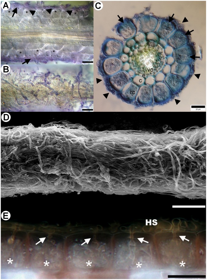
Figure 1. Morphological and anatomical characteristics of sheathed ericoid mycorrhiza from field-collected European blueberry (Vaccinium myrtillus) roots.
1A) Longitudinal section through sheathed ericoid mycorrhiza. The hyphae forming the sheath (arrows) penetrate rhizodermal cell walls (arrowheads) and form dense coils typical for ericoid mycorrhiza (asterisks). DIC, stained with trypan blue, bar = 20 µm. 1B) Surface view of the same sheathed ericoid mycorrhiza displaying the structure of a dense hyphal sheath covering the hair root. DIC, stained with trypan blue, bar = 20 µm. 1C) Cross section of sheathed ericoid mycorrhiza. The hair root is covered by a hyphal sheath (arrowheads), its rhizodermal cells (rc) are filled with dense hyphal coils (arrows). The mycobiont never advances to the cortex/exodermis (c), the endodermis (e) or the vascular cylinder (vc). DIC, stained with trypan blue, bar = 20 µm. 1D) Surface view of sheathed ericoid mycorrhiza showing a dense hyphal sheath. SEM, bar = 50 µm. 1E) Detail of a longitudinal section of sheathed ericoid mycorrhiza. Hyphae forming the hyphal sheath (HS) penetrate rhizodermal cells (arrows) and form coils typical of ericoid mycorrhizal symbiosis (asterisks). DIC, bar = 20 µm.

