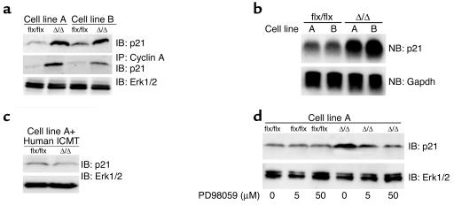
Figure 9.
Increased p21Cip1 protein levels in Icmt-deficient fibroblasts. (a) Extracts from K-Ras-Icmtflx/flx and the derivative K-Ras-IcmtΔ/Δ fibroblasts were analyzed by immunoblotting with an antibody recognizing p21Cip1 (F-5 monoclonal; Santa Cruz Biotechnology Inc.) (upper panel). Cyclin A was immunoprecipitated from the cell extracts with a polyclonal antibody (H-432; Santa Cruz Biotechnology Inc.), and cyclin A–associated p21Cip1 was detected by immunoblotting (middle panel). The blot from the upper panel was stripped and incubated with an anti-Erk1/2 antibody as a loading control (lower panel). Similar results were obtained in three independent experiments. (b) Northern blot of total cellular RNA showing p21Cip1 (Cdkn1a) mRNA levels in K-Ras-Icmtflx/flx fibroblasts and the derivative K-Ras-IcmtΔ/Δ fibroblasts (upper panel). The blot was stripped and probed with a Gapdh cDNA probe as a loading control (lower panel). Similar results were obtained in three independent experiments. (c) Immunoblot showing p21Cip1 protein levels in K-Ras-Icmtflx/flx:ICMT fibroblasts and the derivative K-Ras-IcmtΔ/Δ:ICMT fibroblasts. The blot was stripped and incubated with an anti-Erk1/2 antibody as a loading control (lower panel). (d) K-Ras-Icmtflx/flx and K-Ras-IcmtΔ/Δ fibroblasts were treated overnight with the MEK inhibitor PD98059, and extracts were analyzed by immunoblotting with a p21Cip1-specific antibody. The blot was stripped and incubated with an anti-Erk1/2 antibody as a loading control (lower panel).

