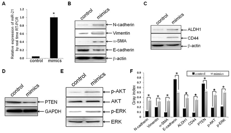
Figure 4. Hsa-miR-21 mimics induced EMT and CSC phenotype, accompanied with PTEN down-regulation and AKT/ERK1/2 activation.
Established MDA-MB-231/anti-miR-21 cells were transfected with hsa-miR-21 mimics at a concentration of 40 nmol for 72 h. (A) MDA-MB-231/anti-miR-21 cells were treated with hsa-miR-21 mimics elevated the expression of miR-21, as compared to control groups (n1 = n2 = 3; p = 0.00373), by real-time RT-PCR analysis. (B-F) Protein levels of mesenchymal markers (N-cadherin, Vimentin and alpha-SMA) (B), epithelial marker (E-cadherin) (B), CSC markers (ALDH1 and CD44) (C), PTEN (D), p-AKT and AKT (E), as well as p-ERK1/2 and ERK1/2 (E) in indicated cells were measured by Western blot analysis, and bands were semi-quantified using ImageJ software (F). Beta-actin or GAPDH was used as loading control. (*indicates p<0.05; ★ indicates p<0.001).

