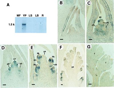
Figure 2.
Distribution of RFL RNA in rice plants. (A) Northern blot analysis. MF, mature florets; YP, young panicles; LS, leaf sheaths; LB, leaf blades; R, roots. (B–G) RFL expression analyzed by in situ hybridization. (B) A vegetative shoot apex. (C and D) A young panicle at primary branch primordia differentiation stage. (E) A young panicle at secondary branch primordia differentiation stage. (F) A developing panicle. Four primary branches at various developmental stages in a panicle are shown. The oldest primary branch (arrow) is in the floret differentiation stage. (G) Developing florets. All floral organis have developed by this stage. pb, primary branch; sb, secondary branch; pa, panicle apex; f, floret. (Bar = 50 μm.)

