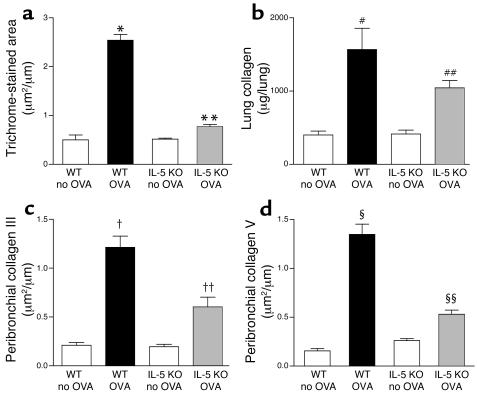
Figure 3.
Quantitation of peribronchial fibrosis in WT and IL-5–deficient mice repetitively challenged with OVA. (a) Area of peribronchial trichrome stain. WT mice repetitively challenged with OVA for 3 months developed an increased area of peribronchial trichrome staining compared with non–OVA-challenged WT mice (*WT OVA versus WT no OVA; P < 0.001). In contrast, IL-5–deficient mice repetitively challenged with OVA for 3 months had significantly reduced areas of peribronchial trichrome staining compared with WT mice repetitively challenged with OVA for 3 months (**IL-5 KO OVA versus WT OVA; P < 0.001). (b) Total lung collagen content. Repetitive OVA challenge induced a significant increase in total lung collagen in WT mice (#WT OVA versus WT no OVA; P < 0.001). IL-5–deficient mice repetitively challenged with OVA had less total lung collagen compared with WT mice repetitively challenged with OVA (##IL-5 KO OVA versus WT OVA; P < 0.05). (c) Collagen III and (d) collagen V lung immunostaining. WT mice repetitively challenged with OVA for 3 months developed increased peribronchial collagen III immunostaining (†WT OVA versus WT no OVA; P < 0.001) (c), as well as increased peribronchial collagen V immunostaining compared with non-OVA–challenged WT mice (§WT OVA versus WT no OVA; P < 0.001) (d). In contrast, IL-5–deficient mice repetitively challenged with OVA had significantly reduced levels of peribronchial collagen III immunostaining (††IL-5 KO OVA versus WT OVA; P < 0.001) (c), as well as significantly reduced levels of peribronchial collagen V immunostaining, compared with WT mice challenged repetitively with OVA for 3 months (§§IL-5 KO OVA versus WT OVA; P < 0.001) (d).

