Figure 4.
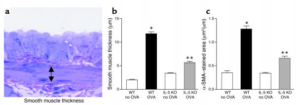
Peribronchial smooth muscle layer in WT and IL-5–deficient mice repetitively challenged with OVA. (a) Peribronchial smooth muscle layer. Lungs that had been fixed in 3% glutaraldehyde and 1% osmium tetroxide were stained with basic fuchsin-toluidine blue, which allowed the best visualization of the peribronchial smooth muscle layer. The thickness of the smooth muscle layer (the transverse diameter) was measured from the innermost aspect to the outermost aspect of the smooth muscle layer (double arrow). (b) Peribronchial smooth muscle layer thickness. WT mice repetitively challenged with OVA for 3 months developed significantly increased thickness of the peribronchial smooth muscle layer compared with non–OVA-challenged WT mice (*WT OVA versus WT no OVA; P < 0.001). In contrast, the thickness of the peribronchial smooth muscle layer in IL-5–deficient mice challenged repetitively with OVA was significantly reduced compared with WT mice challenged repetitively with OVA for 3 months (**IL-5 KO OVA versus WT OVA; P < 0.001). (c) Peribronchial α-smooth muscle actin (α-SMA). WT mice repetitively challenged with OVA for 3 months developed an increase in the area of peribronchial α-smooth muscle actin immunostaining compared with non–OVA-challenged WT mice (*WT OVA versus WT no OVA; P < 0.001). The area of peribronchial α-smooth muscle actin immunostaining in IL-5–deficient mice challenged repetitively with OVA was significantly reduced compared with WT mice challenged repetitively with OVA for 3 months (**IL-5 KO OVA versus WT OVA; P < 0.001).

