
Fig. 1.
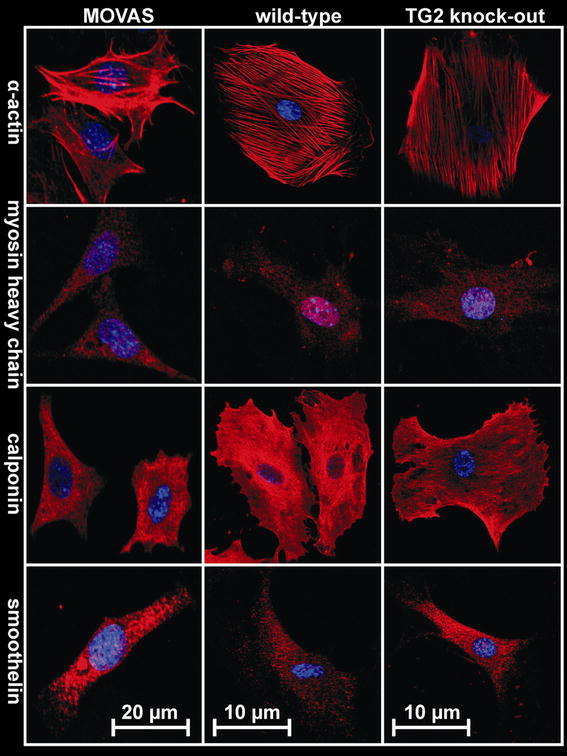
Phenotypical characterization of smooth muscle cells. The MOVAS cell line and explant cells obtained from mesenteric small arteries (WT and KO) were stained for α-actin, myosin heavy chain, calponin and smoothelin, followed by cy-3 secondary antibody (red); nuclei are shown in blue (color figure online)