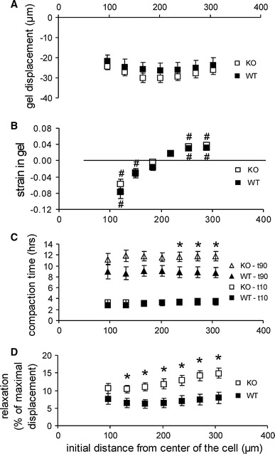
Fig. 3.

Matrix compaction by individual SMCs from wild-type (WT) and TG2 knock-out (KO) mesenteric arteries. a The maximal displacement was not statistically different between WT or KO cells at any measured distance. b Both WT and KO SMCs compacted the matrix at a distance up to 200 μm; between 200 and 300 μm, the matrix expanded locally, while this area moved to the cell center as well. c The time required to achieve the initial 10 % of the maximal displacement was not statistically different between WT and KO cells, but at distances >200 μm the compaction was significantly faster for wild-type SMCs. d Cytochalasin D after 24 h of compaction induced expansion of the matrix due to the loss of cellular contractile forces. The amount of relaxation was significantly lower in WT cells. Asterisk different from zero: P < 0.05 (c, d). Hash symbol WT versus KO: P < 0.05 (b)
