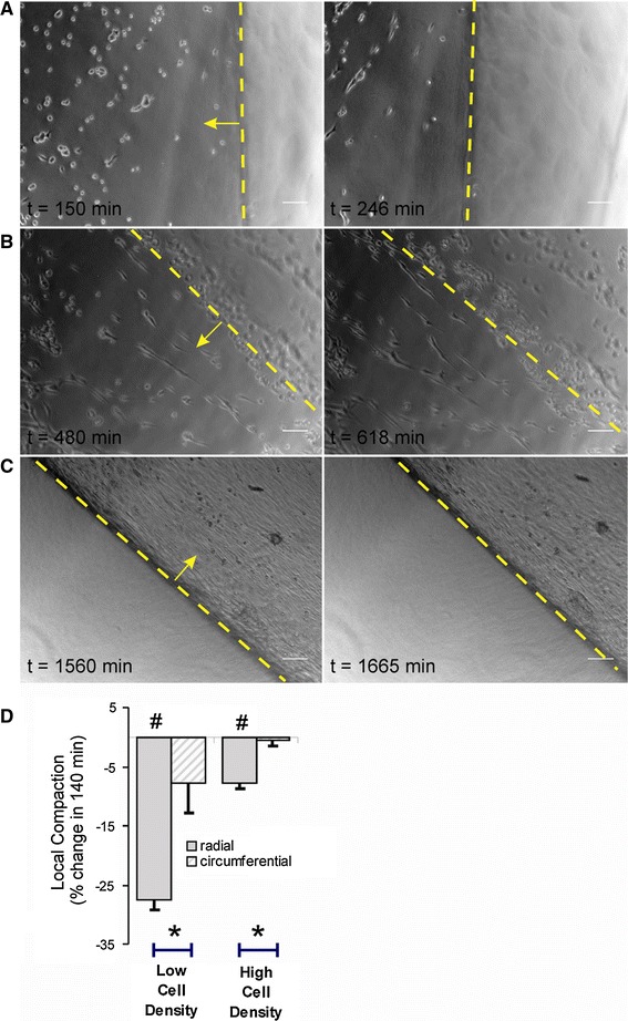
Fig. 8.

Typical cellular orientation during different phases of matrix remodeling as observed near the inner (a, b) and outer (c) border of the ring-shaped collagen gel model (border indicated by a dashed yellow line). a About 2.5 h after cell seeding, cells were still round, but already showed a tendency to align along the inner gel boundary; after 4 h, cells have elongated in this direction, while macroscopic compaction was most visible in the perpendicular direction (indicated by a yellow arrow). b After 8 h, SMC started to develop continuous arrays along the inner boundary, compaction still dominated in the radial direction. c Compaction continued after cells had grown to a confluent layer, this was best visible at the outer gel boundary. d Quantification of a period of 140 min of compaction starting after 480 (“low cell density”, b) and 1,560 min (“high cell density”, c). Local compaction at the boundaries occurred especially in the radial direction of the gel, perpendicular to the cellular long axis, and was high at a low cell density. n = 19, 13, 26, 27. Analyses were limited to cells that occurred in the same image as the gel boundary. Scale bar 100 μm in all panels. Hash symbol different from zero: P < 0.05; asterisk radial versus circumferential: P < 0.05 (color figure online)
