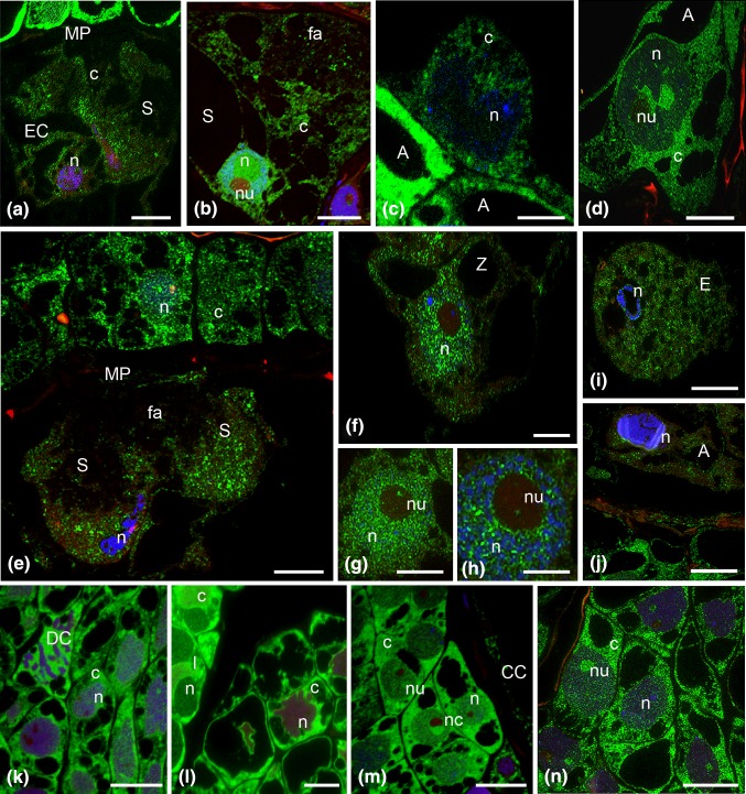
Fig. 2.
Immunofluorescence localization of incorporated 5′-bromouracil of the H. orientalis embryo sac, during the progamic phase: egg cell and synergid cell (a, b), central cell (c), the antipodal cell (d); fertilized embryo sac: degenerated synergid cells with condensed chromatin (e), zygote nucleus (f), the next sections of the zygote nucleus (g, h), endosperm (i), the degenerated antipodal cell (j); somatic cells of the ovule: with 4-nucleate embryo sac (k), with young 7-celled embryo sac (l), during the progamic phase (m) and after fertilization (n). A antipodal cell, c cytoplasm, CC central cell, DC divided cell, E endosperm cell, EC egg cell, fa filiform apparatus, I integument cells, MP micropylar pole, n nucleus, nc nucellus cells, nu nucleolus, S synergid, Z zygote, green newly formed transcripts, blue DAPI staining. Scale bars are 10 μm (a–j) and 20 μm (k–n)

