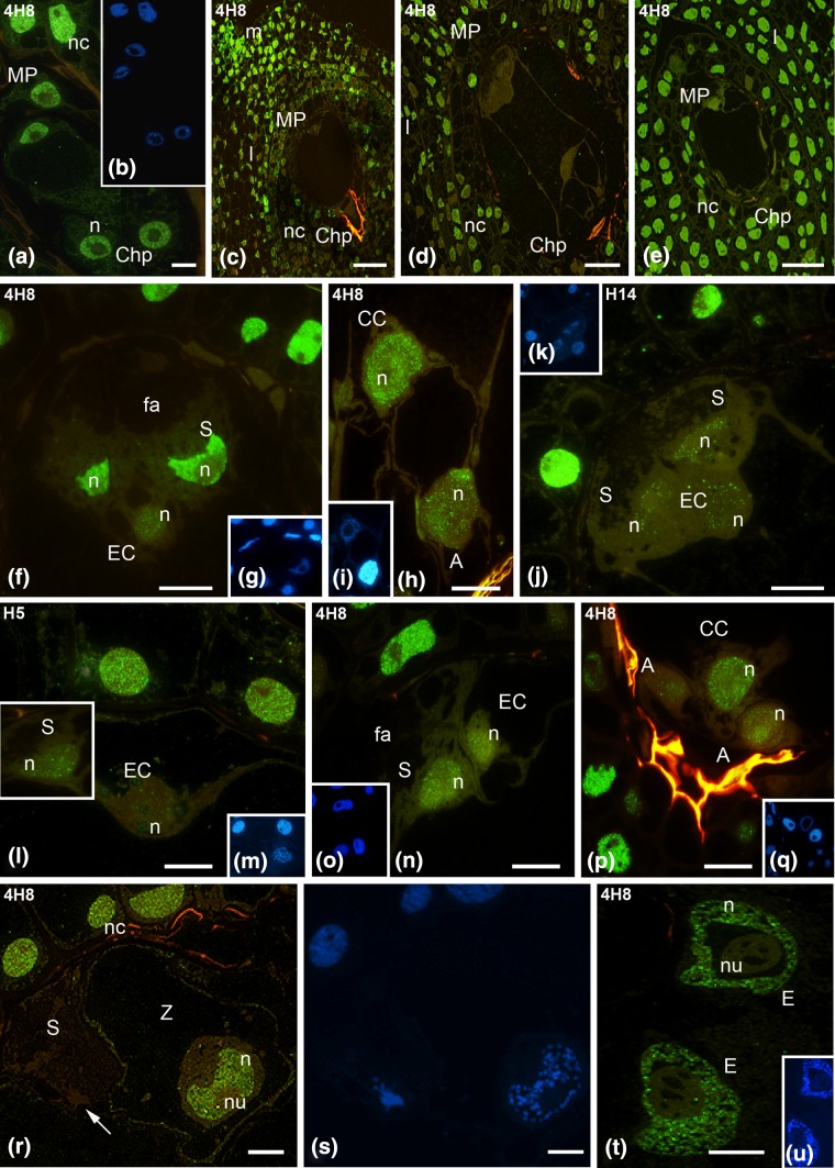
Fig. 3.
Immunofluorescence localization of the total Pol RNA II pool (4H8 antibodies) and the hypophosphorylated (Pol IIA, H14 antibodies) and the hyperphosphorylated (Pol RNA IIO, H5 antibodies) forms of RNA Pol II of the H. orientalis ovule and embryo sac. 4-Nucleate embryo sac (a), mature ovule with the embryo sac after cellularization (c), ovule during the progamic phase (d), ovule after fertilization (e). Mature embryo sac: egg apparatus (f), (h) central and antipodal cells (h), egg apparatus (j–l); embryo sac during the progamic phase: micropylar region (n), chalazal region (p). Embryo sac after fertilization: zygote and degenerated synergid cell (r), endosperm nuclei (t). DAPI staining (b, g, i, k, m, o, q, s, u). m Micropyle, I integument cells, nc nucellus cells, MP micropylar pole, ChP chalazal pole, S synergid, EC egg cell, CC central cell, A antipodal cell, fa filiform apparatus, n nucleus, nu nucleolus, c cytoplasm, green RNA Pol II, blue DAPI staining. Scale bars 10 μm (a, f, h, j–l, n, p, r), 100 μm (c), 50 μm (d, e)

