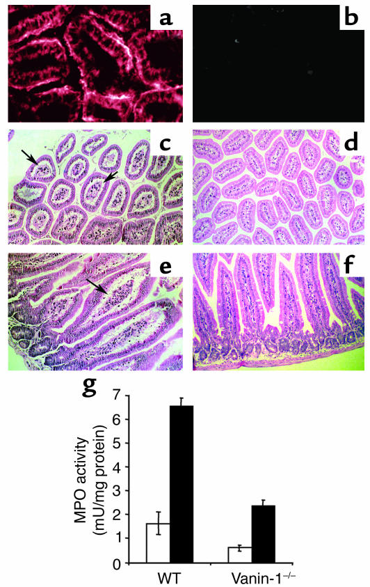
Figure 1.
(a–f) Representative histological sections of small intestine in indomethacin-treated mice. Immunohistology of control WT (a) and Vanin-1–/– (b) mice using anti–Vanin-1 mAb labeling. Cross and longitudinal intestinal sections from WT (c and e) and Vanin-1–/– (d and f) mice; arrows, swollen intestinal villi and dilated lymphatic channels. Hematoxylin-phloxine-saffron staining. Magnification: ×160 (a and b), ×40 (c and d), and ×100 (e and f). (g) Lower intestinal MPO activity in indomethacin-treated WT and Vanin-1–/– mice (white bars, control; black bars, treated). Values are means ± SD. Values from Vanin-1–/– and WT groups are significantly different (P < 0.05, Student t test).

