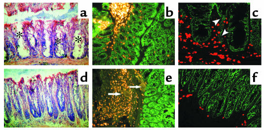
Figure 4.
Cryosections of colon (×100) from three, independent, infected WT (a–c) or Vanin-1–/– (d–f) mice at 8 weeks after infection. Sections were stained with H&E (a and d) or stained with tyramide-Cy3 (b–e), a peroxidase substrate (yellow/red fluorescence) combined with anti-Ep-CAM (green fluorescence) mAb. Note increased mucus production in the crypts of Lieberkühn (*) in WT mice. These pictures are representative of images observed in independent WT (n = 6) or Vanin-1–/– (n = 8) mice, and quantification is performed using the Metamorph program as described in the text. Arrowheads indicate disrupted intestinal mucosa; arrows indicate Schistosoma eggs.

