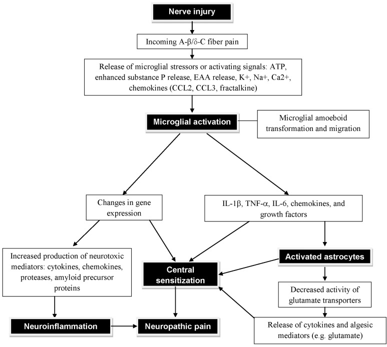
Figure 1.
Diagram showing the neuroinflammation and neuropathic pain subsequent to the neuronal injury and glial cell (microglial and astrocytic) activation. Following a peripheral injury, the synaptic projection of pain sensing neuron within the spinal cord releases ATP. Nearby microglial cells are drawn to the source of ATP and undergo morphological changes as they approach the source and become activated. Fully activated microglial cells are localized around the pain sensing neuron and begin to interact with the neurons at a molecular level, releasing various neuroinflammatory agents. These neuroinflammatory agents activate astrocytes. Upon activation, the astrocytes undergo hypertrophy and increased production of neuroinflammatory agents are secreted into the synaptic cleft. Astrocyte activation in conjugation with microglial activation significantly depolarizes the neuron increasing its sensitivity and potentiating the neuroinflammation and neuropathic pain states. EAA, excitatory amino acids.

