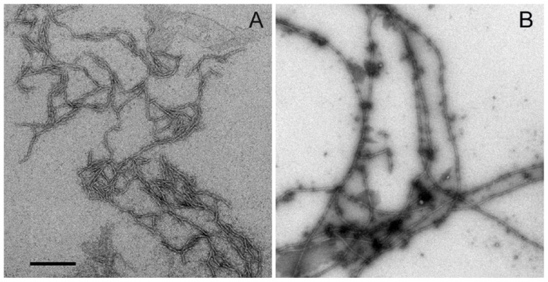
Figure 3.
Negatively stained transmission electron micrographs of Aβ-40 (A) and Aβ-40 Y10F (B). The peptide was diluted in phosphate buffer to 10 μM, and then incubated at 37 °C for 72 h before experiments. Both peptides presented fibrillar morphologies, wild type Aβ-40 showed the combined morphologies of fibrils (5–10 nm in diameter and 400 nm in length) and curvilinear protofibrils (6–8 nm in diameter and <100 nm in length), while Aβ-40 Y10F gave rise to longer mature fibrils (long and extended fibrils >2 μm in length). Scale bar: 200 nm.

